
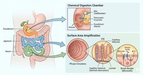
Explore how the duodenum, jejunum, and ileum use villi, peristalsis, and pancreatic enzymes to chemically digest food and absorb vital nutrients.
The Small Intestine: Anatomy, Digestive Functions, and Nutrient Absorption
The small intestine (or small bowel) is the longest and most critical organ of the gastrointestinal tract for nutrient absorption. Following the stomach, it extends approximately 3 to 6 meters (10 to 20 feet) in living adults.
While food is broken down mechanically in the mouth and stomach, the small intestine is where the vast majority of chemical digestion and nutrient extraction takes place.
How the Small Intestine Moves and Cleans Itself
When partially digested food (known as chyme) enters the small intestine from the stomach, the organ utilizes two primary patterns of muscular movement controlled by the enteric nervous system:
- Segmentation Contractions: These localized contractions chop, mix, and roll the chyme back and forth. This ensures the food mixes thoroughly with digestive enzymes and comes into maximum contact with the intestinal wall.
- Peristaltic Contractions: These wave-like contractions slowly propel the chyme forward toward the large intestine.
The Migrating Motor Complex (MMC)
When the small intestine is empty (between meals), a specialized physiological process called the Migrating Motor Complex (MMC) begins. Often referred to as the gastrointestinal "housekeeping wave," these intense cyclical contractions sweep residual food particles, cellular debris, and excess bacteria downward into the large intestine, preventing bacterial overgrowth.
Anatomical Structure: The Three Sections
The small intestine is divided into three distinct, continuous segments, each specialized for specific digestive roles:| Segment | Length | Key Functions & Nutrient Focus |
| Duodenum | ~25 cm (10 inches) | Receives bile and pancreatic juice; primary site for iron absorption and neutralizing stomach acid. |
| Jejunum | ~2.5 meters (8 feet) | Main powerhouse for absorbing carbohydrates (glucose), proteins (amino acids), water-soluble vitamins, and minerals. |
| Ileum | ~2 to 4 meters (6.5 to 13 feet) | Final segment; explicitly responsible for absorbing Vitamin B12, bile salts, and any remaining leftover nutrients. |
1. The Duodenum: The Chemical Mixing Chamber
As the C-shaped duodenum receives acidic chyme, Brunner's glands in the mucosal lining secrete bicarbonate-rich mucus to protect the tissue.
The presence of fats and acids triggers the release of the hormones secretin and cholecystokinin (CCK), which regulate the rate of gastric emptying and stimulate accessory organs:
- The Liver & Gallbladder: Release bile to emulsify large fat globules into tiny droplets.
- The Pancreas: Delivers sodium bicarbonate (to raise pH) and crucial enzymes: lipase (fats), amylase (carbohydrates), and proteases like trypsin (proteins).
2. The Jejunum: The Absorption Engine
Featuring a highly alkaline-to-neutral environment, the jejunum is chemically optimized for massive nutrient transport.
3. The Ileum: The Final Safety Net
The ileum ensures that precious compounds like bile acids are recycled back to the liver (enterohepatic circulation) and captures critical large micronutrients like Vitamin B12 before emptying into the cecum of the large intestine.
Cellular Design: Maximizing Surface Area
The secret behind the small intestine's extraordinary capacity to absorb nutrients lies in its anatomical landscape. The inner lining is modified into three layers of surface amplification that expand its functional area by 30 to 60 times:
- Plicae Circulares: Large, permanent circular folds of the mucosal layer that force chyme to spiral forward rather than move in a straight line.
- Villi: Millions of microscopic, finger-like protrusions (0.5 to 1 mm long) extending into the intestinal lumen. Each villus contains a rich network of blood capillaries and a central lymph vessel (lacteal) to carry absorbed nutrients directly into circulation.
- Microvilli: A dense, microscopic "brush border" coating the surface of each individual villus epithelial cell.
Unlike simple diffusion, most vital nutrients (like amino acids and glucose) are actively transported across this brush border using specialized, energy-requiring cellular channels to ensure maximum extraction.
Frequently Asked Questions (FAQ)
What is the main function of the small intestine?
The primary function of the small intestine is to chemically break down chyme using pancreatic enzymes and bile, and to absorb vitamins, minerals, proteins, carbohydrates, and fats into the bloodstream via microscopic structures called villi.
What are villi and why are they important?
Villi are tiny, finger-like projections lining the small intestinal wall. They drastically increase the internal surface area of the gut, acting as microscopic docks that rapidly capture and transfer nutrients from passing food into the bloodstream.
What happens if the small intestine is damaged or shortened?
Damage to the intestinal lining (as seen in Celiac disease) or surgical removal of a segment can result in malabsorption. Because specific sections absorb specific things, losing parts of the small intestine can lead to severe nutrient deficiencies, chronic diarrhea, and unintended weight loss.
What is the "housekeeping wave" (MMC) in the gut?
The Migrating Motor Complex (MMC) is a pattern of electrical and muscular waves that cleans the stomach and small intestine during fasting states. It pushes remaining food particles and harmful bacteria down into the colon, acting as a crucial defense mechanism against Small Intestinal Bacterial Overgrowth (SIBO).