
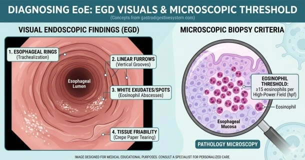
Learn how Eosinophilic Esophagitis (EoE) is diagnosed. Explore what doctors look for during an upper endoscopy (EGD) and understand microscopic biopsy thresholds.
Eosinophilic Esophagitis Diagnosis: What to Expect From Testing
If you experience persistent dysphagia (difficulty swallowing solid foods) or have food frequently feel delayed behind your breastbone, your doctor may suspect Eosinophilic Esophagitis (EoE). Because EoE symptoms closely mimic severe gastroesophageal reflux disease (GERD), structural testing is required to confirm a diagnosis and prevent permanent tissue complications.
The Primary Tool: Upper Endoscopy (EGD)
The definitive process for evaluating the health of your food pipe relies on an Esophagogastroduodenoscopy (EGD), commonly referred to as an upper endoscopy.
During an EGD, a gastroenterologist gently guides a thin, flexible tube equipped with a high-definition camera (an endoscope) down your throat. This allows them to visually examine the delicate inner linings of your esophagus, stomach, and the first part of your small intestine (duodenum) under mild sedation.
Common Endoscopic Findings for EoE
When evaluating a patient with EoE, a specialist typically scans for several distinct structural visual anomalies:
- Esophageal Rings (Trachealization): The presence of numerous concentric rings stacked vertically along the length of the esophagus, making the food pipe look similar to a windpipe.
- Esophageal Strictures: Pronounced narrowing of the esophageal tube that physically restricts the passage of solid foods.
- Linear Furrows: Deep vertical wrinkles, grooves, or channels cutting down the inner layer of the tissue.
- White Exudates: Tiny white spots scattered across the surface lining, which represent microscopic clusters or "mounds" of dying inflammatory eosinophils.
- Tissue Friability (Crepe Paper Esophagus): Esophageal tissue that is exceptionally fragile and easily tears or shears during minor contact with the scope.
The Deciding Factor: Esophageal Biopsy Criteria
While visual abnormalities strongly point toward an EoE diagnosis, an endoscopy alone is not enough to confirm it. An EoE diagnosis must be confirmed through a tissue biopsy.
To perform this, the gastroenterologist passes small, specialized forceps through the endoscope channel to carefully collect minor tissue samples from multiple areas of the esophagus. These samples are then sent to a pathology lab to be analyzed under a microscope.
The Importance of Diagnostic Thresholds
- The 15 Eosinophils Rule: Pathologists look for a specific concentration of inflammatory white blood cells. A definitive diagnosis of EoE requires a peak count of 15 or more eosinophils per high-power field (hpf) within the esophageal tissue.
- Diagnosing a Normal-Looking Esophagus: A biopsy is highly recommended if you suffer from unexplained swallowing difficulties, even if your esophagus looks perfectly healthy during the visual exam. Up to 10-15% of EoE patients present with a visually normal esophagus, yet their microscopic biopsies reveal heavy inflammation.
Secondary Diagnostic Testing Options
While an EGD with biopsies is the absolute clinical standard, doctors may utilize secondary evaluations to map out structural damage or rule out overlapping conditions:
- Barium Swallow (Esophagram): The patient swallows a thick barium liquid while serial X-rays capture its transit. This is exceptionally useful for detecting subtle, long-segment narrowings or strictures that could be missed visually.
- Allergy Testing (Skin Prick or Blood Tests): Though not accurate for identifying exact food triggers inside the esophagus due to EoE's delayed immune pathway, allergy tests help manage concurrent atopic conditions like asthma, eczema, and seasonal hay fever.
Frequently Asked Questions (FAQ)
Is an upper endoscopy painful?
No. An upper endoscopy (EGD) is an outpatient procedure typically performed under conscious sedation or general anesthesia. You will sleep through the test and will not feel the scope or the tissue biopsies being taken. Afterward, some patients experience a mild, temporary sore throat for a day or two.
Can you have EoE if your upper endoscopy looks normal?
Yes. A significant percentage of individuals with EoE have an esophagus that looks completely healthy to the naked eye during an endoscopy. Because of this, doctors must take multiple tissue biopsies from different parts of the food pipe to ensure an accurate diagnosis under the microscope.
How many tissue samples are taken during an EoE biopsy?
Gastroenterologists generally take between 4 to 6 tissue biopsy samples from different areas of the esophagus (typically from both the lower and upper sections). Sampling multiple locations significantly improves diagnostic accuracy and helps ensure that patches of localized inflammation are not missed.