
Learn how doctors diagnose pyloric stenosis. Explore the role of abdominal ultrasound, finding the olive-shaped mass, and identifying electrolyte imbalances.
Pyloric Stenosis Diagnosis: How Do Doctors Confirm It?
Diagnosing pyloric stenosis promptly is crucial to prevent severe dehydration and malnutrition in patients. When an infant presents with classic signs like projectile vomiting, or an adult shows signs of a chronic gastric outlet obstruction, physicians utilize a combination of clinical physical exams, imaging, and lab work to confirm a diagnosis.
The Clinical Physical Examination
A thorough physical exam is often the first step in identifying pyloric sphincter muscle narrowing:
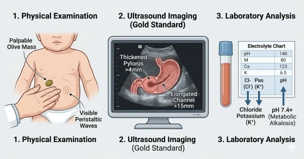
- The "Olive Mass" Palpation: During a physical exam, a pediatric specialist will carefully palpate the infant's right upper abdomen while the baby is relaxed or feeding. In up to 60-80% of cases, the doctor can feel an olive-shaped, firm mass, which is the physically enlarged, hypertrophied pyloric muscle.
- Observation of Peristaltic Waves: The doctor will check the abdomen for visible, wave-like muscle contractions moving from the left upper quadrant across to the right side. This occurs because the stomach is forcefully contracting to squeeze contents past the blockage.
- Hydration Status Assessment: Clinicians check for severe dehydration by evaluating signs like dry mucous membranes, delayed skin turgor, a sunken fontanelle (soft spot), and checking recent weight logs for acute weight loss.
Diagnostic Imaging and Lab Tests
When clinical signs point toward an obstruction, objective imaging and laboratory analysis provide confirmation:
1. Abdominal Ultrasound (The Gold Standard)
An abdominal ultrasound is the definitive imaging tool of choice for infant cases, offering over 95% diagnostic sensitivity and specificity. It uses harmless sound waves to measure the pyloric sphincter muscle dimensions. Ultrasound diagnostic criteria confirming pyloric stenosis typically include:
- A pyloric muscle thickness greater than 3 mm or 4 mm.
- An elongated pyloric channel length greater than 14 mm to 15 mm.
2. Blood Tests and Electrolyte Panels
Frequent vomiting of highly acidic stomach contents (hydrochloric acid) results in a severe, predictable metabolic shift. Blood draws are evaluated for:
- Hypochloremia: Abnormally low chloride levels due to vomiting stomach acid.
- Hypokalemia: Abnormally low potassium levels caused by renal compensation mechanisms.
- Hypochloremic, Hypokalemic Metabolic Alkalosis: A specific shift where the blood's pH rises significantly, becoming too alkaline. These chemistry values must be corrected with IV fluids before any surgery can safely take place.
3. Upper Gastrointestinal (GI) Barium Series
If an ultrasound is inconclusive or if the patient is an adult, a barium swallow test may be performed. The patient drinks a small amount of a chalky liquid (barium) while X-rays track its passage. A classic "string sign" (a thin line of barium trickling through the tightly restricted pylorus) indicates severe narrowing.
Frequently Asked Questions (FAQs)
Why does a doctor look for an "olive-shaped mass"?
The "olive" is actually the physical, thickened pyloric muscle that has grown large due to hypertrophy. Finding this hard mass in the right upper side of the abdomen provides strong clinical evidence of pyloric stenosis before an imaging scan is even performed.
Is an ultrasound safe for a newborn suspected of pyloric stenosis?
Yes, an abdominal ultrasound is completely safe, completely painless, and non-invasive. It uses standard sound waves rather than ionizing radiation, making it the ideal diagnostic choice for infants.
Why do blood tests have to be done before a baby can have surgery?
Because persistent projectile vomiting strips the body of vital acids and essential minerals, babies quickly develop an imbalance called hypochloremic metabolic alkalosis. Surgeons require blood tests to safely ensure these life-threatening imbalances are corrected using intravenous fluids prior to administering general anesthesia.
What is the "string sign" seen on an abdominal X-ray?
The "string sign" is a distinct radiographic visual clue seen during a barium swallow test. It shows up when the thick, chalky barium contrast fluid is squeezed into a tiny, thread-like stream as it struggles to pass through the severely narrowed pyloric channel.