
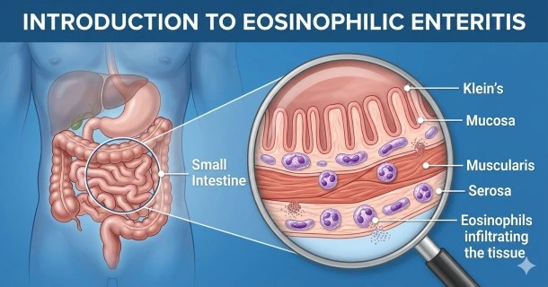
Learn about eosinophilic enteritis, a rare chronic inflammatory disorder where eosinophils infiltrate the small intestine wall, disrupting normal digestion.
What is Eosinophilic Enteritis? An Introduction to a Rare Gastrointestinal Disorder
Eosinophilic enteritis is a rare, chronic inflammatory condition characterized by the abnormal accumulation of eosinophils—a specific type of white blood cell—within the tissue layers of the small intestine. It belongs to a broader family of immune-mediated conditions known as Eosinophilic Gastrointestinal Diseases (EGIDs). Under normal physiological conditions, eosinophils play a minor role in fighting off parasitic infections and regulating allergic reactions. However, in patients with eosinophilic enteritis, these cells flood the small intestine without an active infection, releasing toxic granules that cause tissue inflammation, swelling, and structural dysfunction.
Because the small bowel is responsible for the vast majority of nutrient absorption, chronic inflammation in this region can severely disrupt systemic health if left unmanaged.
Anatomical Mapping: Understanding Where EGIDs Strike
Eosinophilic gastrointestinal disorders are named strictly according to the specific segment of the digestive tract where the eosinophilic infiltration occurs:
- Eosinophilic Esophagitis (EoE): Confined strictly to the esophagus.
- Eosinophilic Gastritis (EG): Isolated to the mucosal lining of the stomach.
- Eosinophilic Enteritis (EE): Target inflammation occurs primarily inside the small intestine (duodenum, jejunum, or ileum).
- Eosinophilic Colitis (EC): Affects the large intestine (colon).
Pathophysiology: Klein’s Structural Classification
The clinical presentation and impact of eosinophilic enteritis depend heavily on which specific layer of the small intestinal wall the eosinophils infiltrate. According to the widely accepted Klein Classification, the disease presents in three main structural forms:
- Mucosal Layer Involvement: This is the most common presentation. Eosinophils infiltrate the innermost lining (mucosa), causing patchy ulcerations and blunting the microscopic intestinal villi. This layer damage leads directly to malabsorption, protein-losing enteropathy, and chronic diarrhea.
- Muscularis Layer Involvement: The white blood cells penetrate the middle muscular layers of the small bowel. This causes marked thickening and rigidity of the intestinal wall, frequently leading to localized bowel obstructions, severe abdominal cramping, and vomiting.
- Serosal Layer Involvement: The rarest and most distinct form, where eosinophils cross into the outer lining (serosa). This typically presents with pronounced fluid accumulation in the abdomen, a clinical complication known as eosinophilic ascites.
Key Epidemiological & Diagnostic Realities
- Prevalence: Eosinophilic enteritis is an exceptionally rare disease, estimated to affect fewer than 1 in 100,000 individuals globally, though diagnostic rates are climbing due to increased clinical awareness.
- Bimodal Age Groups: While it can affect anyone at any age, it is most frequently diagnosed in children and young adults, often showing a slight male predominance.
- The Atopic Connection: A significant majority of individuals diagnosed with this condition have a concurrent history of allergic diseases, such as asthma, eczema, allergic rhinitis, or severe food IgE-mediated sensitivities.
- Diagnostic Markers: Confirming the condition requires a combination of high peripheral blood eosinophil counts and a formal upper endoscopy or capsule endoscopy with multiple tissue biopsies showing dense, abnormally high clusters of eosinophils per high-power field (HPF).
Frequently Asked Questions (FAQs)
Is eosinophilic enteritis a type of inflammatory bowel disease (IBD)?
No, eosinophilic enteritis is not a form of Inflammatory Bowel Disease (like Crohn’s disease or ulcerative colitis). While both conditions cause chronic inflammation in the digestive tract, their underlying biological pathways are completely distinct. IBD features generalized autoimmune and bacterial-driven tissue damage, whereas eosinophilic enteritis is driven specifically by allergic-type white blood cell infiltration and eosinophilic degranulation.
Can eosinophilic enteritis be cured with dietary changes alone?
While dietary elimination strategies are highly effective, eosinophilic enteritis is considered a chronic, lifelong condition rather than something easily cured. For many patients, identifying and removing specific food antigens (such as dairy, wheat, or soy) via a Six-Food Elimination Diet (SFED) or utilizing an elemental formula diet can induce long-term remission. However, severe cases often require supplemental medical treatments like topical or systemic corticosteroids.
What happens if eosinophilic enteritis is left untreated?
Without appropriate clinical monitoring and treatment, untreated eosinophilic enteritis can progress to severe structural bowel complications. Chronic muscular layer inflammation can lead to recurrent bowel obstructions requiring emergency surgery, while deep mucosal damage can cause severe nutritional wasting, profound iron-deficiency anemia, and childhood growth failure.