
Learn how specialists diagnose eosinophilic enteritis. Discover the essential role of endoscopies, biopsies, fluid analysis, and ruling out other diseases.
How is Eosinophilic Enteritis Diagnosed? A Guide to Medical Testing and Criteria
Diagnosing eosinophilic enteritis can be a complex clinical challenge. Because its symptoms mimic more common conditions like celiac disease, inflammatory bowel disease (IBD), and standard food allergies, gastroenterologists diagnose eosinophilic enteritis using a strict process of elimination combined with microscopic analysis of tissue biopsies.
To secure a definitive diagnosis, a patient must meet specific clinical criteria: the presence of gastrointestinal symptoms, histological evidence of high eosinophil counts in the small bowel, and the total exclusion of other systemic or parasitic causes of eosinophilia.
1. Endoscopic Evaluation and Tissue Biopsies
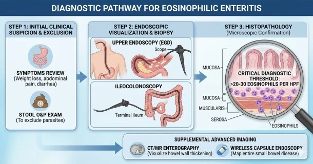
The absolute gold standard for confirming eosinophilic enteritis is an endoscopic look inside the small bowel, coupled with tissue harvesting.
- Upper Endoscopy (EGD) and Enteroscopy: A gastroenterologist passes a thin, flexible camera down the esophagus, through the stomach, and deep into the small intestine (duodenum and jejunum). Doctors look for signs like mucosal swelling, white patches, patchy ulcerations, or a loss of normal intestinal folds.
- Multiple Tissue Biopsies: Because eosinophilic infiltration is frequently patchy, doctors must take multiple tissue samples from both normal and abnormal-looking areas of the small intestine.
- Microscopic Thresholds (Histopathology): A pathologist examines these tissue samples under a microscope. While a few eosinophils can normally reside in the gut, finding more than 20 to 30 eosinophils per high-power field (HPF) in the small bowel tissue provides the definitive structural proof needed for diagnosis.
2. Advanced Diagnostic Imaging and Capsule Endoscopy
When inflammation sits deep within the middle or lower segments of the small bowel, standard scopes cannot reach it. Advanced imaging bridges this gap:
- Wireless Capsule Endoscopy: The patient swallows a small, pill-sized camera capsule that takes thousands of images as it moves naturally through the digestive tract. This is highly effective for mapping mucosal disease throughout the entire length of the small intestine.
- CT or MR Enterography: These specialized cross-sectional scans use oral contrast fluid to expand the small intestine. They are essential for evaluating patients with suspected muscularis layer involvement, as they clearly visualize bowel wall thickening, rigid strictures, or partial obstructions.
3. Laboratory Screening and Fluid Analysis
Blood, stool, and fluid tests are used to support the biopsy findings and systematically rule out competing diagnoses.
- Complete Blood Count (CBC) with Differential: This blood test checks for peripheral eosinophilia—an elevated count of eosinophils circulating in the bloodstream. While present in many patients, a normal blood count does not rule out the disease, as the inflammation can be entirely localized to the gut tissue.
- Stool Ova and Parasite (O&P) Exam: Because parasitic infections are a primary cause of high eosinophil counts, doctors must run multiple stool tests to completely rule out active parasites before diagnosing an immune disorder.
- Paracentesis (for Serosal Disease): If a patient has a fluid-filled abdomen (ascites) from suspected serosal layer disease, a doctor will draw a fluid sample using a needle. Finding massive concentrations of eosinophils in this fluid confirms serosal eosinophilic enteritis.
4. Allergy Testing and Dietary Evaluation
Once the intestinal damage is proven to be eosinophilic, allergy assessments help pinpoint the underlying external drivers.
- Skin Prick and Serum IgE Tests: These standard allergy tests check for immediate hypersensitivities to common foods (like milk, wheat, soy, and eggs). However, because eosinophilic enteritis involves a delayed, non-IgE cell-mediated immune response, these tests only provide a partial map and are used alongside elimination diets.
Frequently Asked Questions (FAQs)
Can a standard colonoscopy or endoscopy miss eosinophilic enteritis?
Yes, a standard endoscopy or colonoscopy can miss the condition if tissue biopsies are not taken or if the disease is patchy. If a doctor takes samples from only one healthy-looking spot, or if the inflammation is trapped in the middle of the small intestine out of reach of standard scopes, the disease can be overlooked. This is why taking multiple biopsies and using capsule endoscopies or scans is crucial.
What is the difference between an allergy test and a biopsy for this diagnosis?
An allergy test identifies potential external substances that might provoke your immune system, whereas a biopsy provides physical proof of actual structural tissue damage inside your body. Allergy tests cannot diagnose eosinophilic enteritis on their own; only a microscopic count of eosinophils from a tissue biopsy can confirm the condition.
What other diseases must be ruled out before confirming eosinophilic enteritis?
Doctors must systematically rule out intestinal parasites, celiac disease, Crohn’s disease, hypereosinophilic syndrome (HES), and gastrointestinal cancers. Because all of these conditions can cause overlapping symptoms or elevated eosinophil levels, a diagnosis of eosinophilic enteritis is only official when these alternative causes are fully investigated and excluded.