
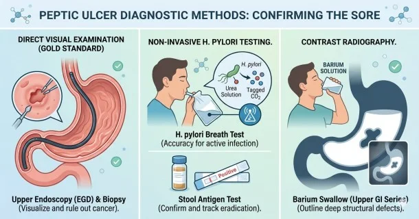
Discover how doctors test for and confirm peptic ulcers. Compare the gold-standard upper endoscopy against non-invasive H. pylori breath and stool antigen tests.
Peptic Ulcer Diagnosis: Common Medical Tests and Confirmation
Confirming a diagnosis of peptic ulcer disease (PUD) is crucial for preventing severe upper gastrointestinal complications. While a healthcare provider can strongly suspect an ulcer based on characteristic patterns of epigastric pain, diagnostic validation is required to differentiate an ulcer from severe gastritis, GERD, or gastric malignancies, and to select the correct targeted medical therapy.
Depending on your overall health status, age, and clinical presentation, a gastroenterologist will deploy structural imaging techniques or highly specific pathogen screenings.
1. Direct Visual Examination (The Gold Standard)
Direct structural evaluation allows a specialist to visually confirm the existence, size, and precise location of a mucosal tear.
- Upper Endoscopy (EGD): An esophagogastroduodenoscopy is the definitive gold standard for confirming a peptic ulcer. A specialist passes a thin, flexible tube equipped with a high-definition camera down the esophagus, into the stomach, and through the duodenum. Unlike older clinical assumptions, endoscopy is highly practical as it allows doctors to visually map the ulcer, grade its severity, and immediately treat active bleeding sites.
- Endoscopic Tissue Biopsy: During an endoscopy, specialized tools pass through the tube to gather miniature samples of the stomach lining. A pathologist checks these cells under a microscope, providing the most accurate method to definitively rule out stomach cancer and verify localized bacterial colonies.
2. Targeted Screenings for H. pylori Bacteria
Because Helicobacter pylori bacteria drive up to 90% of duodenal ulcers, identifying this pathogen is a key step in a diagnostic workup. Non-invasive screenings are heavily favored for initial validation:
- Urea Breath Test (UBT): An exceptionally accurate, non-invasive diagnostic test. You will ingest a baseline solution containing tagged carbon molecules. If H. pylori is present, its urease enzymes break down the urea solution into ammonia and carbon dioxide, allowing the tagged carbon to be precisely measured when you blow into a collection bag.
- Stool Antigen Test: This highly reliable lab screening detects distinct viral or bacterial proteins (antigens) shed by H. pylori directly into the feces. It is widely used by clinicians to confirm an initial active infection and to verify complete eradication a few weeks after antibiotic therapy.
- H. pylori Serology (Blood Test): An inexpensive screening that measures circulating antibodies against the bacteria. While easy to perform, it is less clinically useful because antibodies can remain positive for months or years after an infection has been completely cured, making it unable to distinguish between a past exposure and an active ulcer trigger.
3. Contrast Radiography
- Barium X-Rays (Upper GI Series): A traditional imaging method where you drink a thick, chalky barium liquid that temporarily coats the inner lining of the upper digestive tract. A series of X-rays are then taken, utilizing the barium silhouette to highlight large structural abnormalities, structural niches, or deep mucosal craters along the stomach and intestinal walls. While useful, it lacks the ability to collect tissue biopsies or detect flat, early-stage erosions.
Frequently Asked Questions (FAQ)
Is an upper endoscopy necessary for everyone with stomach ulcer symptoms?
Not always, but it is heavily recommended for individuals over the age of 60 or those displaying critical "alarm signs" such as unintentional weight loss, difficulty swallowing, persistent vomiting, or signs of internal bleeding. For younger patients with mild symptoms, a doctor may choose to perform a non-invasive H. pylori screening first before moving to an endoscopy.
Why can a blood test give a false positive for an active H. pylori infection?
An H. pylori blood test measures the presence of specific antibodies generated by your immune system. Because these protective antibodies can remain in your bloodstream for several years after the bacteria have been completely eliminated by antibiotics, the test cannot accurately differentiate between a past, fully healed exposure and a current, active ulcer-causing infection.
What makes the stool antigen test so highly recommended for follow-up care?
The stool antigen test only yields a positive result when active, living H. pylori proteins are being shed through the digestive tract. This makes it an exceptional, highly accurate tool for doctors to use four to six weeks after a patient finishes their antibiotic regimen to guarantee that the infection has been completely eradicated.