
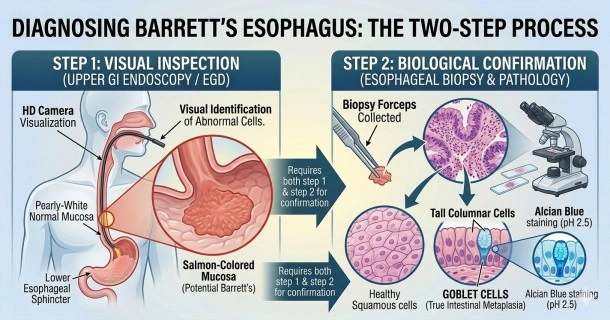
Learn how Barrett's esophagus is diagnosed using upper GI endoscopy and histological biopsy analysis to identify specialized intestinal metaplasia.
Barrett's Esophagus Diagnosis: Clinical Testing and Evaluation
Because the cellular transformation of the food pipe is frequently asymptomatic, a definitive Barrett's esophagus diagnosis cannot be made based on physical symptoms alone. Clinical guidelines recommend that adults with a long history of chronic Gastroesophageal Reflux Disease (GERD)—particularly those with multiple risk factors—undergo specialized screening. Confirming the condition requires an upper gastrointestinal evaluation combined with tissue sampling.
The Primary Screening: Upper GI Endoscopy (EGD)
An upper GI endoscopy procedure (also known as an esophagogastroduodenoscopy or EGD) is the primary diagnostic tool used to visualize the health of the upper digestive tract.
Patient Preparation and Safety
To ensure clear visualization and maximum safety during the procedure, patients must follow strict preparation guidelines:
- NPO (Fasting) Guidelines: Patients must not eat or drink anything for 4 to 8 hours before the procedure. Chewing gum and smoking are also restricted, as they stimulate gastric juices.
- Medication Review: Patients must inform their gastroenterologist of all current medications. Certain drugs may need to be temporarily paused or adjusted under medical supervision:
- Blood thinners (anticoagulants and antiplatelets like aspirin, warfarin, or clopidogrel).
- Nonsteroidal anti-inflammatory drugs (NSAIDs like ibuprofen or naproxen).
- Certain blood pressure, diabetes, or antidepressant medications that might interact with clinical sedatives.
Visualizing the Tissue Shift
During an endoscopy, a thin, flexible tube equipped with a high-definition camera and light is passed through the mouth down into the esophagus, stomach, and duodenum. The physician carefully inspects the tissue lining:
- Normal Esophageal Mucosa: Displays a pale, pearly-white color composed of stratified squamous cells.
- Metaplastic Barrett's Mucosa: Appears as a distinct, velvety salmon-colored mucosa extending upward from the gastroesophageal junction.
While a salmon-colored appearance strongly suggests tissue modification, visual inspection alone is insufficient. The tissue must be pathologically verified under a microscope.
Pathological Confirmation via Esophageal Biopsy
During the endoscopy, the physician passes miniature forceps through a channel in the scope to perform an esophageal biopsy. Small tissue samples are collected from the salmon-colored areas, following structured protocols to map the extent of the tissue shift.
Histological Analysis and Goblet Cells
The tissue samples are sent to a pathology lab where they are analyzed under a microscope to distinguish between different types of cell structures:
- Gastric Metaplasia: Cells that resemble the lining of the stomach. This indicates irritation but does not confirm a diagnosis of Barrett's esophagus.
- Specialized Intestinal Metaplasia: True Barrett's tissue characterized by the clear presence of goblet cells in the esophagus. Goblet cells are large, mucus-secreting cells normally found exclusively in the intestines. Their presence is the definitive diagnostic marker required by international medical guidelines to confirm Barrett's esophagus.
Pathologists frequently use specialized clinical stains, such as Alcian Blue (pH 2.5), to clearly highlight the acid mucins within goblet cells, distinguishing true intestinal metaplasia from other structural mimics. The pathology report will also classify the sample to check for any signs of pre-cancerous mutations (dysplasia).
Frequently Asked Questions (FAQs)
How is Barrett's esophagus officially confirmed?
Barrett's esophagus is officially confirmed using a two-step process: an upper endoscopy and an esophageal biopsy. The doctor must visually identify salmon-colored tissue in the lower esophagus, and a pathologist must find specialized intestinal goblet cells under a microscope.
Does an upper endoscopy for Barrett's esophagus hurt?
No, the procedure itself is not painful. Patients are typically given a local anesthetic spray to numb the throat and suppress the gag reflex, alongside an intravenous sedative to maximize comfort and help the patient relax or sleep during the short evaluation.
What does "salmon-colored mucosa" mean in an endoscopy report?
Salmon-colored mucosa refers to a deep pink or reddish tissue lining seen in the lower food pipe. In a healthy esophagus, the lining is pale and pearly-white. A shift to a salmon color indicates that the cells have changed and require a biopsy to check for Barrett's esophagus.