
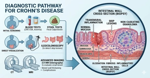
Learn how gastroenterologists diagnose Crohn's disease. Explore endoscopy, colonoscopy, imaging, blood markers, and biopsy criteria used for a definitive diagnosis.
How is Crohn’s Disease Diagnosed? A Guide to Medical Tests and Procedures
Diagnosing Crohn’s disease requires a comprehensive approach because no single test can definitively identify the condition. Gastroenterologists use a differential diagnosis process, combining blood work, advanced imaging, and endoscopic evaluations to rule out other gastrointestinal disorders and visually confirm deep tissue inflammation.
Getting a timely, accurate diagnosis is the first critical step toward establishing an effective management plan and avoiding long-term complications like bowel strictures or fistulas.
1. Initial Laboratory Screenings (Blood and Stool Tests)
Before performing invasive procedures, doctors run laboratory tests to look for systemic signs of inflammation, anemia, and nutritional deficiencies.
- Inflammatory Markers (CRP and ESR): Blood tests measuring C-Reactive Protein (CRP) and Erythrocyte Sedimentation Rate (ESR) detect active, generalized inflammation throughout the body.
- Complete Blood Count (CBC): A CBC screens for anemia (low red blood cell count), which often results from chronic internal intestinal bleeding or poor iron absorption.
- Fecal Calprotectin Test: This highly sensitive stool test detects calprotectin, a protein released by white blood cells in the intestines. High levels provide clear evidence of localized intestinal inflammation, helping doctors differentiate between active inflammatory bowel disease (IBD) and non-inflammatory issues like Irritable Bowel Syndrome (IBS).
2. Endoscopic Evaluations (Direct Visualization)
Endoscopy is the gold standard for diagnosing Crohn’s disease, allowing physicians to visually inspect the mucosal tissue lining the digestive tract.
- Ileocolonoscopy: A flexible tube with a camera is guided through the entire length of the colon and into the terminal ileum (the final section of the small intestine). This allows doctors to identify the classic "skip lesions" (patchy areas of inflammation separated by healthy tissue) and the irregular "cobblestone" mucosal patterns characteristic of Crohn's.
- Upper GI Endoscopy: If a patient experiences upper abdominal symptoms, an endoscopy is performed to look for inflammation or ulcerations in the esophagus, stomach, and duodenum.
- Capsule Endoscopy: For cases where inflammation is isolated deep within the small intestine—an area out of reach for traditional scopes—the patient swallows a small, pill-sized wireless camera that transmits thousands of images as it travels naturally through the digestive tract.
3. Histopathology (Tissue Biopsies)
During an endoscopic procedure, a gastroenterologist will collect small tissue samples (biopsies) from both inflamed and healthy-looking sections of the bowel.
A pathologist examines these micro-samples under a microscope to confirm the diagnosis. The presence of transmural inflammation (cellular damage cutting deep through multiple tissue layers) and non-caseating granulomas (organized clusters of immune cells) provides definitive histopathological confirmation of Crohn's disease.
4. Advanced Diagnostic Imaging
Radiological imaging helps determine the exact extent of the disease, mapping out deep tissue involvement and identifying hidden structural complications.
- CT or MR Enterography: These specialized scans require the patient to drink a contrast solution that highlights the small intestine. They are highly effective at detecting bowel wall thickening, strictures (narrowed pathways), obstructions, or deep fluid collections (abscesses).
- Pelvic MRI: This imaging method is highly accurate for evaluating the perianal region, mapping out complex fistula tracts or deep abscesses near the rectum.
Frequently Asked Questions (FAQs)
Can a colonoscopy miss Crohn’s disease?
Yes, a standard colonoscopy can occasionally miss Crohn's disease if the inflammation is entirely restricted to the upper portions of the small intestine, which a colonoscope cannot physically reach. If your doctor suspects small bowel involvement despite a normal colonoscopy, they will typically order a capsule endoscopy, a CT enterography, or an MRI to evaluate the hidden areas.
What is the difference between a Crohn's diagnosis and an Ulcerative Colitis diagnosis?
The distinction relies entirely on the location and depth of the inflammation found during testing. Crohn's disease can appear anywhere along the GI tract (from mouth to anus), leaves healthy "skip" patches between inflamed areas, and cuts deep through all layers of the tissue. Ulcerative Colitis is confined strictly to the colon and rectum, presenting with continuous inflammation that only impacts the innermost mucosal layer.
How long does it typically take to get a definitive Crohn's diagnosis?
The diagnostic process can take anywhere from a few weeks to several months. Because early symptoms like abdominal cramping and loose stools mimic common issues like food intolerances, infectious gastroenteritis, or IBS, doctors must methodically rule out these conditions through sequential blood, stool, and imaging tests before confirming an IBD diagnosis.