
How do doctors diagnose tropical sprue? Learn about diagnostic criteria, blood panels, malabsorption tracking, and endoscopic small bowel biopsy requirements.
How is Tropical Sprue Diagnosed? Clinical Criteria and Medical Tests
The diagnosis of Tropical Sprue is established through a strict process of clinical exclusion, requiring confirmed evidence of multi-nutrient malabsorption alongside structural small intestinal tissue abnormalities. Because its symptoms closely mirror common parasitic infections, inflammatory bowel diseases, and Celiac disease, a definitive diagnosis can only be made after ruling out alternative causes.
A complete diagnostic workup evaluates an individual's travel history, nutrient levels in the blood, and small bowel mucosal tissue architecture.
Step 1: Evaluating Clinical History and Risk Anchors
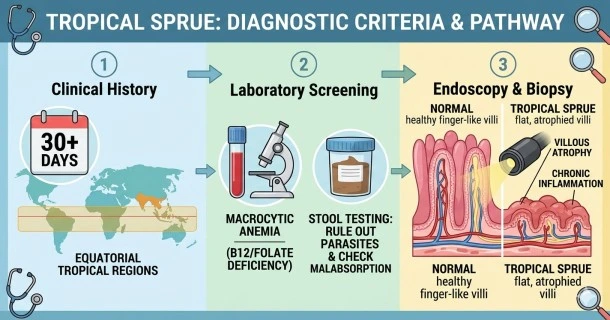
Before ordering invasive diagnostic procedures, a healthcare provider will verify if a patient meets the baseline environmental exposure requirements:
- Geographic Exposure: Proof of extended travel or residence (typically lasting more than 30 consecutive days) within a known tropical or subtropical endemic zone.
- Symptom Duration: Persistent gastrointestinal complications and soft or greasy stools that have continuously failed to resolve for several weeks or months.
Step 2: Comprehensive Laboratory Screening
Once risk exposure is confirmed, an array of blood and stool tests is executed to detect metabolic signs of small intestinal failure:
- Complete Blood Count (CBC): Used to identify macrocytic/megaloblastic anemia. This presents as abnormally large red blood cells, which directly indicates a systemic lack of folic acid and vitamin B12.
- Serum Nutrient Panels: Blood draws explicitly measure active circulating concentrations of Vitamin B12, Serum Folate, Iron, and Vitamin D to assess the baseline severity of malabsorption.
- Stool Microbiological Testing: Comprehensive PCR assays, microscopy, and antigen tests are ordered to explicitly rule out persistent parasitic or bacterial pathogens, such as Giardia, Cryptosporidium, and Entamoeba histolytica.
- Quantitative Fecal Fat Collection: A 24-hour to 72-hour stool evaluation tracking fat absorption. Excreting more than 6 to 7 grams of fat per day confirms significant fat malabsorption (steatorrhea).
- D-Xylose Absorption Test: An oral sugar absorption test. Low excretion levels of D-xylose in a patient's urine or blood indicate that the proximal mucosa of the small intestine is damaged and failing to absorb carbohydrates properly.
Step 3: Endoscopy and Histological Biopsy (The Gold Standard)
To confirm the structural layout of the disease, an Esophagogastroduodenoscopy (EGD) is scheduled. A gastroenterologist passes a flexible camera down into the first portions of the small intestine to inspect the mucosa and gather tiny tissue samples (biopsies).
| Diagnostic Modality | Normal Presentation | Tropical Sprue Histology |
|---|---|---|
| Endoscopic Visuals | Smooth, distinct pink mucosal folds in the duodenum. | Scalloping, flattening, or blunt cracking of the duodenal mucosal rings. |
| Villi Architecture | Long, finger-like projections maximizing absorption space. | Subtotal or total villous atrophy; the villi appear flattened and blunt. |
| Crypt Anatomy | Shallow cellular depressions between adjacent intestinal villi. | Marked crypt elongation or hyperplasia as the body attempts to generate new cells. |
| Immune Cell Infiltration | Minimal leukocyte presence inside the tissue layers. | Profound infiltration of intraepithelial lymphocytes throughout the epithelial sheets. |
Differential Diagnosis: What Must Be Ruled Out?
To fulfill diagnostic criteria, a physician must run targeted medical tests to differentiate Tropical Sprue from clinical lookalikes:
- Celiac Disease Exclusions: Blood serology tests tracking Tissue Transglutaminase Antibodies (tTG-IgA/IgG) and Anti-Endomysial Antibodies (EMA) are drawn. These tests are consistently negative in cases of Tropical Sprue. Additionally, a Tropical Sprue biopsy often reveals severe damage extending deep into the distal ileum, whereas Celiac disease classically diminishes in severity further down the small bowel.
- Crohn’s Disease Exclusions: Colonoscopies or cross-sectional imaging check for chronic deep transmural ulcers, granulomas, or structural strictures, which do not occur in uncomplicated Tropical Sprue.
Frequently Asked Questions (FAQs)
Can an endoscopy alone diagnose Tropical Sprue?
No. While an endoscopy can physically reveal flattened intestinal folds and a biopsy can confirm villous atrophy, these microscopic findings look nearly identical to Celiac disease or severe microbial overgrowth. A definitive diagnosis requires correlating the biopsy results with a history of tropical exposure and ruling out celiac antibodies.
What is the single most defining factor in confirming a Tropical Sprue diagnosis?
Because it is a diagnostic process of exclusion, the ultimate confirmation often rests on a positive clinical response to therapeutic trial treatment. If a patient’s chronic diarrhea stops and their small bowel lining completely heals following a combination course of tetracycline antibiotics and folic acid supplements, the diagnosis is confirmed.
Why do doctors look for megaloblastic anemia when testing for this condition?
Megaloblastic anemia is a direct biochemical indicator that the middle and lower sections of the small intestine (the jejunum and ileum) have suffered severe mucosal damage. This injury prevents the absorption of folic acid and vitamin B12, halting normal red blood cell development and signaling advanced malabsorption.
Is a breath test used during a Tropical Sprue diagnostic workup?
Hydrogen breath tests, which are routinely utilized to diagnose standard Small Intestinal Bacterial Overgrowth (SIBO), may be integrated into selected cases. However, because Tropical Sprue presents with more severe, structural villous destruction and systemic macrocytic anemia than standard SIBO, a breath test is considered a secondary assessment rather than a standalone diagnostic tool.