
Discover how celiac disease is accurately diagnosed. Learn about tTG-IgA blood tests, endoscopic small bowel biopsies, genetic testing, and the mandatory gluten challenge timeline.
Celiac Disease Diagnosis: Clinical Guidelines, Blood Tests & Biopsy Protocols
An accurate celiac disease diagnosis requires a structured, multi-step medical evaluation combining serological blood tests, genetic screening, and a confirmatory small bowel biopsy. Because the treatment involves a strict, lifelong lifestyle change, ruling out look-alike conditions via standardized diagnostic criteria is essential.
Crucially, all diagnostic testing for celiac disease must be performed while you are still consuming a gluten-containing diet. Eliminating gluten prematurely can cause blood antibody levels to drop and the intestinal lining to heal, leading to false-negative test results.
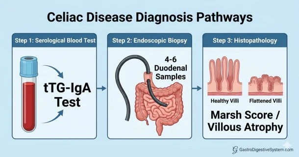
1. First-Line Screening: Serological Blood Testing
The initial step in identifying celiac disease involves detecting specific autoimmune antibodies in the blood.
- tTG-IgA (Tissue Transglutaminase IgA Antibody): This is the premier screening tool due to its high diagnostic accuracy. It exhibits a sensitivity and specificity exceeding 95% when performed on individuals who regularly ingest gluten.
- Total Serum IgA Level: This must be checked concurrently with the tTG-IgA test. Approximately 2% to 3% of patients with celiac disease suffer from selective IgA deficiency, which can result in false-negative tTG-IgA readings.
- DGP-IgG (Deamidated Gliadin Peptide IgG Antibody): This alternative marker is heavily relied upon if a patient presents with an underlying selective IgA deficiency, or during evaluations for infants and toddlers under the age of two.
- EMA-IgA (Endomysial Antibody IgA): Possessing a specificity near 100%, this test is frequently deployed as a secondary, confirmatory serological assay to verify borderline or highly elevated tTG-IgA levels.
2. The Gold Standard: Small Bowel Endoscopic Biopsy
If celiac serology yields a positive result, an esophagogastroduodenoscopy (EGD) with a small bowel biopsy is standard practice to confirm architectural damage.
- The Procedure: A gastroenterologist passes a flexible endoscope through the stomach into the proximal part of the small intestine (the duodenum) to collect small tissue specimens.
- Sampling Requirements: Guidelines mandate collecting a minimum of 4 to 6 tissue biopsies (including at least 1 or 2 from the duodenal bulb and 4 from the distal duodenum) due to the patchiness of celiac lesions.
- Histopathological Evaluation (The Marsh Score): Pathologists examine the samples under a microscope to classify the severity of tissue changes:
- Marsh 1: Normal villous architecture but a significant influx of intra-epithelial lymphocytes (IELs).
- Marsh 2: Crypt hyperplasia along with elevated intra-epithelial lymphocytes.
- Marsh 3: True villous atrophy, where the microscopic, finger-like projections (villi) responsible for nutrient absorption are flattened, blunted, or entirely absent.
3. The Evolving No-Biopsy Diagnostic Pathway
Historically restricted to pediatric cases, modern international medical guidelines recognize a selective no-biopsy celiac diagnosis pathway for adult criteria under strict parameters:
| Diagnostic Prerequisite | Clinical Threshold Requirements |
|---|---|
| High-Titer Serology | Initial tTG-IgA levels must exceed \(\ge \) 10 times the Upper Limit of Normal (ULN) on a standard assay. |
| Secondary Verification | A separate, independent blood draw must confirm the presence of EMA-IgA antibodies. |
| Symptomatic Presentation | The patient typically presents with classic malabsorptive or high-risk systemic symptoms. |
Note: If the tTG-IgA value is elevated but falls below 10 times the upper limit of normal, a confirmatory endoscopic biopsy remains mandatory.
4. Navigating Prior Gluten Avoidance: The Gluten Challenge
A major hurdle in modern gastroenterology clinics is evaluating individuals who have already initiated a gluten-free diet without undergoing prior diagnostic evaluations. Because the immune-mediated damage subsides without dietary gluten exposure, baseline serology and biopsies will appear normal, causing false-negative outcomes.
- The Diagnostic Protocol: A structured gluten challenge is necessary before blood tracking or endoscopic procedures can yield definitive results.
- Duration and Intake: Patients are instructed to consume approximately 3 to 10 grams of gluten daily (roughly equivalent to 2 slices of standard wheat bread) for a minimum of 2 to 6 weeks.
- Clinical Monitoring: Serological tracking is typically conducted at the conclusion of the challenge, followed by an upper endoscopy if antibodies rise or symptoms flare significantly.
Frequently Asked Questions (FAQs)
What is the accuracy of celiac disease serology blood tests?
When a patient consumes a normal, gluten-containing diet, first-line blood screening via the tTG-IgA assay demonstrates exceptional accuracy, yielding a sensitivity and specificity greater than 95%. However, accuracy drops drastically if gluten has been eliminated prior to the blood draw.
What does it mean if my tTG-IgA test is right at the upper limit of normal?
Borderline elevations near the upper limit of normal require cautious interpretation. Your gastroenterologist will likely order a secondary, highly specific Endomysial Antibody (EMA-IgA) test or schedule a small bowel biopsy to verify if low-grade active inflammation or early enteropathy is occurring.
Can an adult be diagnosed with celiac disease without an upper endoscopy biopsy?
Yes, under strict criteria. Updated clinical guidelines permit a no-biopsy diagnosis in adults if the initial tTG-IgA blood test registers extremely high—specifically at or above 10 times the laboratory's standard upper limit of normal—and a secondary, independent blood sample tests positive for EMA-IgA antibodies.
How long do I need to eat gluten for a gluten challenge to be accurate?
To ensure diagnostic reliability, a standard gluten challenge requires consuming 2 slices of gluten-containing bread daily for a minimum of 2 to 6 weeks. Short-term challenges lasting under 2 weeks risk missing tissue abnormalities in up to 10% of individuals.