
Learn how specialists diagnose and stage stomach cancer. Explore endoscopy biopsy findings, EUS depth measurement, and TNM staging criteria.
Stomach Cancer Diagnosis and Staging
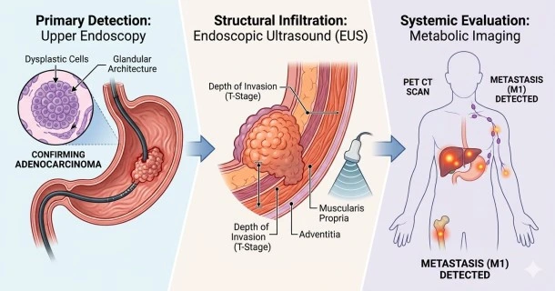
Stomach cancer diagnosis relies primarily on an upper endoscopy combined with tissue biopsy to identify malignant cells. This complex process confirms the presence of gastric adenocarcinoma. It also determines the tumor's exact location. Once confirmed, staging tests evaluate how far the disease has progressed.
Primary Diagnostic Procedures
Gastroenterologists use highly targeted procedures to view the stomach lining and collect tissue samples.
- Upper Endoscopy (EGD): Direct visualization of the inner mucosal surface. It identifies ulcerated masses or abnormal tissue rigidness.
- Endoscopic Biopsy: Extraction of multiple suspicious tissue samples. Pathologists analyze these cells under a microscope.
- Endoscopic Ultrasound (EUS): Evaluation of endoscopic ultrasound gastric wall depth. It determines if tumors have invaded the muscular layers. It also assesses nearby lymph nodes.
Advanced Staging and Tracking Modalities
Oncologists use advanced imaging and laboratory tests to map the spread of the disease.
- CT Scan: Detailed cross-sectional imaging of the abdomen. It reveals local tumor extension into adjacent organs.
- PET CT Scan: High-resolution metabolic imaging. It detects hidden PET CT scan stomach cancer metastasis in distant regions.
- Diagnostic Laparoscopy: Minimally invasive surgical inspection of the abdomen. It checks for tiny tumor implants on the peritoneum.
- Liquid Biopsy: Non-invasive blood testing. It enables liquid biopsy stomach cancer tracking by analyzing circulating tumor DNA (ctDNA).
Gastric Cancer Staging Criteria (TNM)
The staging system categorizes the tumor based on anatomical progression. Medical teams utilize the American Joint Committee on Cancer (AJCC) framework.
| Stage Component | Clinical Focus | Structural Milestones |
|---|---|---|
| T (Tumor) | Depth of structural invasion | Ranges from T1 (inner mucosa) to T4 (outer serosa layer). |
| N (Nodes) | Regional lymph node involvement | Ranges from N0 (no nodes) to N3 (16 or more positive nodes). |
| M (Metastasis) | Distant spread through the body | Classified as M0 (no distant spread) or M1 (active distant metastasis). |
Frequently Asked Questions
What specific tissue markers are evaluated during a stomach cancer biopsy?
Pathologists test the tissue for specific genetic and protein expressions. They routinely screen for HER2 protein amplification. They also evaluate PD-L1 expression levels and Mismatch Repair (MMR) status. These biomarkers dictate whether targeted biological therapies or immunotherapies will be effective.
Why is a diagnostic laparoscopy performed before stomach cancer surgery?
CT and PET scans frequently miss microscopic clusters of cancer cells on the abdominal lining. A diagnostic laparoscopy allows the surgeon to physically look inside the peritoneal cavity with a camera. Finding these tiny cellular implants changes the stage to M1, preventing unnecessary major stomach surgery.
What do the stomach cancer endoscopy biopsy findings look like?
Malignancies usually present in one of three ways during an endoscopy. They can look like a deep, irregular ulcer with raised borders. They may appear as a distinct, bulky, projecting mass. In diffuse cases, they can show up as a flat, thick, rigid section of the stomach wall.