
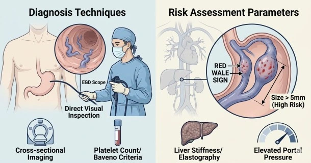
How are stomach varices diagnosed? Learn about upper endoscopy, non-invasive Baveno criteria, and key bleeding risk factors identified during screening.
Stomach Varices Diagnosis and Risk Assessment
Stomach varices diagnosis relies on clinical screening protocols to locate enlarged gastric veins before they cause dangerous internal bleeding. Because these blood vessels develop silently beneath the stomach lining due to Portal Hypertension, proactive screening is mandatory for patients with chronic liver diseases. Modern medical protocols use a mix of direct imaging and non-invasive scoring systems to evaluate a patient's variceal rupture risk factor profile.
How Doctors Diagnose Stomach Varices
Gastroenterologists utilize several specialized diagnostic tests outlined in clinical literature like StatPearls to locate and classify gastric varices:
- Upper Endoscopy (EGD): This is the gold standard diagnostic procedure. A thin, lighted tube with a camera is guided down the esophagus into the stomach. It allows doctors to see the exact size, location, and condition of the veins.
- Endoscopic Ultrasound (EUS): This advanced tool combines endoscopy with ultrasound waves. It is highly effective for evaluating deep fundic varices that might be missed during a standard EGD.
- Contrast-Enhanced CT or MRI Scans: Cross-sectional imaging provides a comprehensive map of the abdominal blood vessels. It is especially useful for diagnosing isolated stomach varices caused by splenic vein blood clots.
- Doppler Ultrasound: A non-invasive imaging scan used to evaluate the speed and direction of blood flow through the portal venous system.
Non-Invasive Screening: Avoiding Unnecessary Endoscopies
According to the Baveno criteria highlighted by the European Society of Gastrointestinal Endoscopy (ESGE), certain low-risk patients with compensated cirrhosis can safely defer an invasive endoscopy. An EGD can often be delayed if the patient meets both of the following non-invasive markers:
- Platelet Count: Higher than 150 × 10⁹/L.
- Transient Elastography (Liver Stiffness): Measures lower than 20 kPa.
If a patient's platelets drop below this threshold or liver stiffness increases, a diagnostic endoscopy must be scheduled promptly.
Critical Risk Factors Evaluated During Diagnosis
During a routine diagnostic screening, a physician does not just check for the presence of varices. They actively grade specific structural indicators to determine the patient's immediate risk of a life-threatening hemorrhage:
- Variceal Size: Veins exceeding 5 mm in width carry a substantially higher risk of structural wall failure and rupture.
- The "Red Wale" Sign: The appearance of cherry-red streaks or spots on the surface of the vein. This indicates the vessel wall is dangerously thin and prone to bursting.
- Sarin Classification Type: Identifying whether the varices are connected to the esophagus (GOV1/GOV2) or completely isolated in the stomach fundus (IGV1). Isolated fundic varices carry a significantly higher bleeding severity risk.
- Child-Pugh Severity Score: Patients classified with advanced liver failure (Class C) have compromised blood-clotting functions, which elevates their clinical bleeding risk profile.
Frequently Asked Questions (FAQs)
How often do you need to be screened for stomach varices?
For patients diagnosed with liver cirrhosis, baseline screening is recommended immediately. If no varices are detected, standard guidelines suggest repeating an upper endoscopy every 2 to 3 years. If small, stable varices are found, screening intervals shift to every 1 to 2 years to monitor for progression.
Is an upper endoscopy painful?
No. An upper endoscopy (EGD) is an outpatient procedure typically performed under light conscious sedation or general anesthesia. Patients sleep through the brief examination and generally feel no pain, though a mild sore throat may persist for a day.
Can a standard abdominal ultrasound detect stomach varices?
A standard abdominal ultrasound can show indirect signs of portal hypertension, such as an enlarged spleen or fluid accumulation (ascites). However, it cannot reliably view the detailed blood vessels inside the stomach lining. Direct endoscopic evaluation or a contrast-enhanced CT scan is required for a definitive diagnosis.