
Learn how specialists diagnose Ménétrier's disease. Discover key endoscopic findings, deep biopsy protocols, lab tests, and long-term cancer risk screening.
Diagnosing Ménétrier’s Disease
Diagnosing Ménétrier's disease requires a combination of endoscopic visualization, deep tissue biopsies, and specialized laboratory evaluations. Because this rare hyperplastic gastropathy closely mimics other structural stomach conditions, precise cellular confirmation is vital. The diagnostic process focuses heavily on identifying giant cerebriform gastric folds while ruling out malignancy and alternative protein-wasting disorders.
Primary Diagnostic Procedures
Confirming a diagnosis involves a step-by-step clinical evaluation to visualize the stomach lining and extract tissue samples.
- Upper Endoscopy (EGD): Evaluates the physical structure of the stomach wall. It reveals massive, brain-like mucosal folds that typically affect the body and fundus while sparing the antrum.
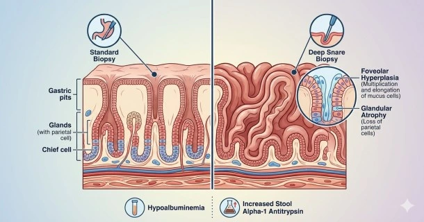
- Deep Mucosal Biopsy: Extracts deep tissue samples using specialized snare techniques. Standard superficial biopsies often fail because they miss the deep structural modifications.
- Endoscopic Ultrasound (EUS): Measures the specific layers of the stomach wall. It confirms that the thickening is confined to the mucosal layer, helping rule out deep wall infiltration.
Key Lab and Pathological Findings
Pathologists and gastroenterologists look for a highly specific triad of chemical and cellular anomalies:
Histopathology (Tissue Analysis)
- Foveolar Hyperplasia: Extreme elongation and corkscrew twisting of mucus-producing surface cells.
- Glandular Atrophy: Prominent loss of specialized acid-producing parietal cells and chief cells.
- Mucous Metaplasia: Replacement of healthy gastric glands with mucus-secreting tissue.
Laboratory Data
- Hypoalbuminemia: Severely depleted blood albumin levels caused by continuous gastric leaking.
- Gastric pH Testing: High pH readings confirming low stomach acid production (achlorhydria).
- Alpha-1 Antitrypsin Clearance: Elevated stool markers confirming active protein-losing enteropathy.
Differential Diagnosis
Several gastrointestinal conditions present with thickened stomach folds. Specialists use the following distinctions to confirm Ménétrier’s disease:
| Condition | Primary Distinguishing Feature | Acid Secretion Status |
|---|---|---|
| Ménétrier’s Disease | Foveolar hyperplasia with marked protein loss | Markedly decreased (Achlorhydria) |
| Zollinger-Ellison Syndrome | Gastrin-secreting tumor causing rugal growth | Massively increased |
| Gastric Lymphoma / MALT | Malignant infiltration of white blood cells in stomach wall | Variable |
| Hypertrophic Gastritis | Severe, active inflammatory cell infiltration | Normal to variable |
Evaluating Long-Term Risks and Surveillance
Once diagnosed, the clinical focus shifts toward assessing ongoing complications and structural risks:
- Infection Screening: Adults are routinely screened for H. pylori bacteria, while pediatric cases are tested for active Cytomegalovirus (CMV).
- Malignancy Monitoring: Because adult-onset Ménétrier’s disease is a premalignant condition, it carries an increased risk of developing gastric adenocarcinoma.
- Surveillance Protocols: Patients must undergo scheduled, periodic upper endoscopies with multiple surveillance biopsies to detect early signs of dysplasia or cancerous transformations.
- Once diagnostic confirmation is established, immediate therapeutic intervention is required. Learn about dietary adjustments, biological therapies, and surgical options in our guide to Ménétrier’s Disease Treatment.
Frequently Asked Questions
Why is a standard stomach biopsy sometimes insufficient to diagnose Ménétrier's disease?
Standard endoscopic biopsies only shave off the very top layer of the stomach lining (mucosa). In Ménétrier’s disease, the characteristic changes—such as glandular atrophy and deep foveolar elongation—occur deep within the gastric pits. A superficial biopsy may mistake the tissue for a simple hyperplastic polyp, making a deeper snare or full-thickness mucosal biopsy necessary for accuracy.
Can an imaging scan like a CT or MRI replace an endoscopy for diagnosis?
No. While a CT scan or MRI of the abdomen can readily show abnormally thickened stomach walls or giant folds, these imaging modalities cannot view the mucosal surface directly nor harvest tissue. They are useful secondary tools to rule out outside masses, but an endoscopy with histopathological tissue analysis remains the gold standard.
What risk factors prompt a doctor to screen a Ménétrier’s patient for stomach cancer?
The primary risk factor is the chronic cellular turnover and tissue alterations (metaplasia) inherent to the adult form of the disease. If surveillance biopsies reveal epithelial dysplasia (precancerous cell changes), or if the patient develops unexplained iron-deficiency anemia or fecal occult blood, the frequency of endoscopic cancer screening is immediately increased.